Author: adminKR
Bioinformatika Konsep Dasar Protein dan Penelusuran Sekuen Protein
Prototipe Krim Ekstrak Uncaria gambir
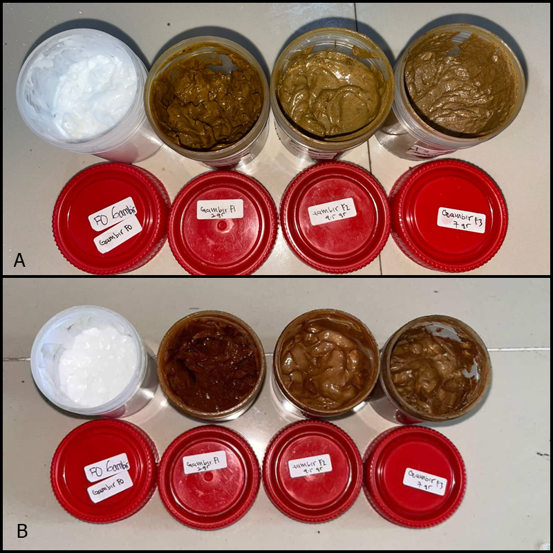
Krim ekstrak gambir (Uncaria gambir) merupakan sediaan topikal berbahan alami yang dikembangkan dari ekstrak etanol gambir yang kaya akan senyawa katekin dan polifenol. Senyawa aktif tersebut diketahui memiliki aktivitas antiinflamasi, antioksidan, dan berpotensi membantu mempercepat proses perbaikan jaringan kulit. Pengembangan prototipe krim ini diarahkan sebagai alternatif terapi topikal berbasis bahan alam yang lebih aman dan memiliki potensi aplikasi pada gangguan kulit inflamasi seperti psoriasis.
Prototipe produk krim ekstrak gambir ini telah dipublikasikan pada jurnal Current Applied Science and Technology dalam kategori online first dengan judul: “Activities Analysis of Uncaria gambir (U. gambir) Ethanol Extract in Vaseline-based Cream to Alleviate Psoriasis in Imiquimod-induced Rats”. Penelitian tersebut menunjukkan bahwa formulasi krim ekstrak gambir mampu menurunkan derajat keparahan psoriasis pada model hewan coba, memiliki stabilitas fisik yang baik, serta tidak menunjukkan efek sistemik yang signifikan.
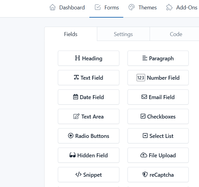
KR Formulir
Aplikasi ini untuk membuat formulir sesuai keperluan penelitian atau studi.
Link aplikasi klik di sini
Pengumuman Mahasiswa Terpilih Anggota Riset Dosen Hibah Dikti 26
Buka pengumuman menggunakan Laptop / PC
Mahasiswa Terpilih Anggota Riset Dosen Hibah Dikti 26
| Nama | Prodi | Hasil | Calon Pembimbing 1 | Ketua Hibah |
|---|---|---|---|---|
| Rika Syaharani | D4 Teknologi Laboratorium Medis | Diterima | Dr. Khoirul RA, Ns. M. Biomed | Dr. Khoirul RA, Ns. M. Biomed |
| Fera Amelia | D4 Teknologi Laboratorium Medis | Diterima | Dr. Khoirul RA, Ns. M. Biomed | Dr. Khoirul RA, Ns. M. Biomed |
| Duta Ardika Saputra | D4 Teknologi Laboratorium Medis | Diterima | Fath Dwisari, M.Si | Fath Dwisari, M.Si |
| Sintya Kusumaningrum | D4 Teknologi Laboratorium Medis | Diterima | Fath Dwisari, M.Si | Fath Dwisari, M.Si |
| Awlya Aryana | D4 Teknologi Laboratorium Medis | Diterima | Ariffialdi Nurhidayattulloh, S.Tr., M. Kes | Dr. Khoirul RA, Ns. M. Biomed |
| Pelagia Abzel Dea | D4 Teknologi Laboratorium Medis | Diterima | Natasya Intan Ramadhani, S.Tr., M.Kes | Natasya Intan Ramadhani, S.Tr., M.Kes |
| Rhabiyatul adawiyah | D4 Teknologi Laboratorium Medis | Diterima | Natasya Intan Ramadhani, S.Tr., M.Kes | Natasya Intan Ramadhani, S.Tr., M.Kes |
| Lisa Safitri | D4 Teknologi Laboratorium Medis | Diterima | Ariffialdi Nurhidayattulloh, S.Tr., M. Kes | Dr. Khoirul RA, Ns. M. Biomed |
| Fina Aulia Shafira | D4 Teknologi Laboratorium Medis | Tidak diterima | ||
| Tea kristiani | D4 Teknologi Laboratorium Medis | Tidak diterima | ||
| Elsa Idrus | D4 Teknologi Laboratorium Medis | Tidak diterima | ||
| Hernicha Febriani | D4 Teknologi Laboratorium Medis | Tidak diterima | ||
| Serli Wahyuni | D4 Teknologi Laboratorium Medis | Tidak diterima | ||
| Riska Auliasari | D4 Teknologi Laboratorium Medis | Tidak diterima | ||
| Puji Nurmayanti | D4 Teknologi Laboratorium Medis | Tidak diterima | ||
| Nabila Oktavia Ramadhani Ayu Wulandari | D4 Teknologi Laboratorium Medis | Tidak diterima | ||
| alpin | D4 Teknologi Laboratorium Medis | Tidak diterima | ||
| SUCI RAHMAH WATI | D4 Teknologi Laboratorium Medis | Tidak diterima | ||
| Nazwa azahra | D4 Teknologi Laboratorium Medis | Tidak diterima | ||
| Dea Ananda Putri | D4 Teknologi Laboratorium Medis | Tidak diterima | ||
| SYF SHEISA SAHARA | D4 Teknologi Laboratorium Medis | Tidak diterima | ||
| DESSY SUYATMI | D4 Teknologi Laboratorium Medis | Tidak diterima | ||
| Alya annisa | D4 Teknologi Laboratorium Medis | Tidak diterima | ||
| Peri irawan | D4 Teknologi Laboratorium Medis | Tidak diterima | ||
| DAVINA MERDEKA PUTRI | D4 Teknologi Laboratorium Medis | Tidak diterima | ||
| ummi kulsum | D4 Teknologi Laboratorium Medis | Tidak diterima |
Mahasiswa yang terpilih dapat menghubungi calon pembimbing 1 yang tertera dalam tabel untuk informasi berikutnya.
Aplikasi Android Pola Asuh Sehat
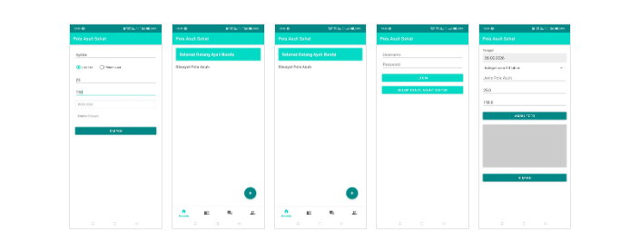
Aplikasi ini diinisiasi oleh dosen prodi kebidanan poltekkes kemenkes Pontianak. Aplikasi Pola Asuh Sehat merupakan luaran hasil penelitian yang ditujukan untuk membantu ibu-ibu dalam memberikan pola asuh yang tepat bagi anak mereka. Fitur utama aplikasi ini diantaranya:
1. Tambah data anak
2. Analisis tumbuh kembang anak
3. Tambah foto pola asuh anak
4. Alarm pengingat pola asuh
Aplikasi ini dapat didownload di Google play store, klik di https://play.google.com/store/apps/details?id=com.polaasuhsehat
KR Exambro – Aplikasi Pencegah Kecurangan Ujian
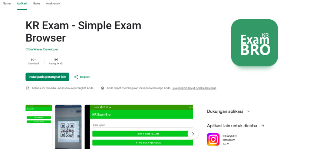
Aplikasi ini dirancang untuk mencegah kecurangan saat berlangsung ujian menggunakan perangkat android. Dengan aplikasi ini, maka peserta ujian tidak bisa:
1. beralih ke layar lain sebelum dia menyelesaikan ujian
2. Menerima notif selama ujian
Fitur yang tersedia:
1. masuk link ujian
2. masuk ke website ujian menggunakan scan QR
Aplikasi ini telah tersedia di Google Playstore klik di sini
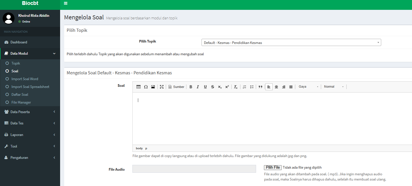
CBT Kr Center

CBT ini merupakan CBT yang bisa digunakan dosen atau guru untuk memberikan latihan kuis saat memberikan pengajaran. CBT ini dilengkapi fitur:
1. Membuat soal dan pilihan jawaban (pilihan ganda, essay)
2. Fitur waktu kuis
3. Fitur jawab dan penilaian otomatis
4. Token
5. Kartu peserta
6. dan masih banyak fitur lainnya
Apabila bapak / ibu tertarik untuk menggunakan CBT dengan custom sesuai nama sendiri, Bapak / Ibu dapat berlangganan CBT ini melalui formulir yang saya sediakan di website ini.
Biaya langganan Rp 500.000 / tahun sudah memperoleh subdomain cbt sesuai nama Bapak / Ibu. Akses halaman admin ke web CBT.